CARTILAGE INJURIES · ABU DHABI

Cartilage injury changes the joint surface.The decision depends on where it is—and what your knee needs to do.

Articular cartilage creates the smooth, low-friction surface of the knee. A focal defect may follow injury or occur alongside meniscal or ligament damage, and its importance depends on symptoms, swelling, function, lesion characteristics and the condition of the rest of the joint. Prof. Dr. Sammy Hanna connects those pieces before discussing rehabilitation, joint-preservation strategies or surgery. Cartilage injury treatment in Abu Dhabi is available at Healthpoint Hospital.

Active adult descending outdoor steps with controlled knee loading

CHAPTER 01 · THE PATTERN

What does the knee do under load?

Choose the closest description. These patterns help organise the assessment; they do not diagnose a cartilage defect on their own.

PATTERN 01AN EDUCATIONAL GUIDE

Pain builds with loaded movement, impact or deeper flexion.

A symptomatic cartilage lesion may become more noticeable when the joint repeatedly carries load. The examination asks where the pain is felt, what movements provoke it and whether swelling follows activity.

See what the examination checks

CHAPTER 02 · EXAMINATION

Test how the knee moves, loads and controls the joint—not just where it hurts.

Cartilage symptoms are not specific enough to diagnose from pain alone. The examination connects the symptom pattern with swelling, range, alignment, control, joint-line or patellofemoral findings and any instability.

  1. 01

    What movement actually reproduces the problem?

    Walking, stairs, squatting, running, jumping and change of direction load different parts of the knee and help define the functional pattern.

  2. 02

    Is there swelling or loss of motion?

    Effusion and restricted flexion or extension suggest intra-articular irritation and help determine whether the knee needs further investigation.

  3. 03

    Where does the load appear to concentrate?

    Alignment, patellar tracking, single-leg control and the location of tenderness provide context for which compartment or surface may be symptomatic.

  4. 04

    Are the meniscus and ligaments intact?

    Cartilage injury can occur alongside meniscal or ligament injury, so stability and meniscal signs should be assessed rather than assuming one isolated diagnosis.

  5. 05

    What has changed in real life?

    Sport, work, stairs, kneeling, walking distance, swelling after activity and confidence under load matter more than an imaging grade alone.

FUNCTION / 02
Clinician observing a patient during a controlled single-leg functional knee assessment
“A useful assessment asks how the joint behaves under load—and whether the cartilage finding explains that behaviour.”

CHAPTER 03 · IMAGING

MRI can define the joint surface.It still cannot choose the treatment by itself.

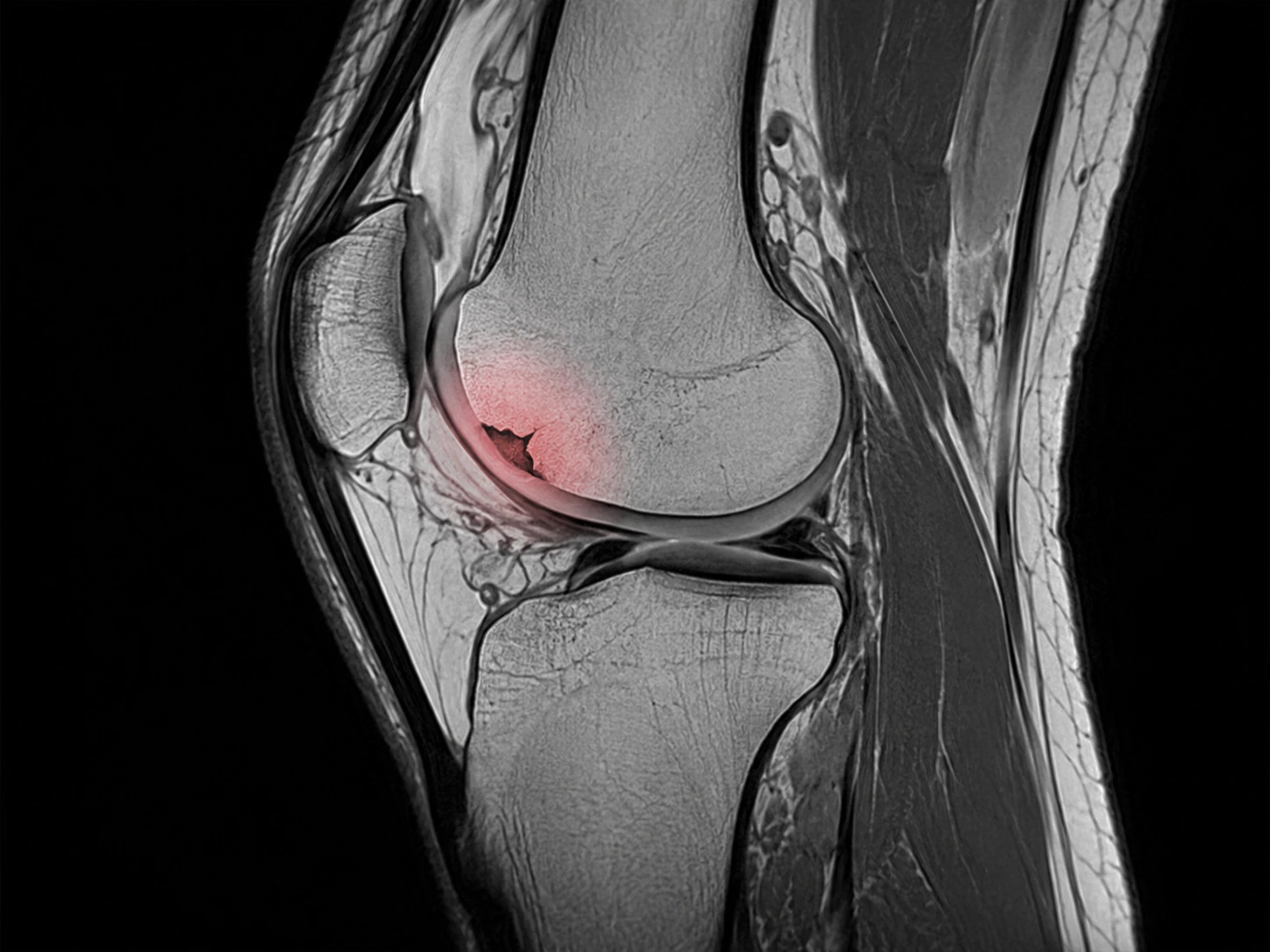
Plain X-rays help assess alignment and established arthritis but do not directly show articular cartilage. MRI can help define a focal defect and evaluate the underlying bone, menisci, ligaments and the rest of the joint when that information will change the plan.

Educational MRI-style sagittal knee illustration showing a highlighted focal articular cartilage defect
Educational MRI-style illustrationNot patient-specific

SELECTED OBSERVATION 01 / 03

Look at the articular surface first

A focal chondral lesion is defined by loss, fissuring or irregularity of the articular cartilage surface. MRI interpretation considers the exact site and depth rather than treating “cartilage damage” as one uniform diagnosis.

CHAPTER 04 · OPTIONS

Treatment follows the clinical problem.
Protect the joint before chasing the scan.

The route depends on symptoms, lesion size and depth, location, underlying bone, alignment, meniscal and ligament status, previous care, activity demands and the condition of the remaining joint surface.

ROUTE01

BUILD CAPACITY

Improve what the knee can tolerate under load.

What it may include

Education, temporary load modification, progressive quadriceps and hip strengthening, mobility work and graded return to the activities that matter.

What it cannot promise

Rehabilitation does not regrow normal hyaline cartilage, but it can improve strength, confidence, load tolerance and symptoms while clarifying whether the lesion is clinically important.

See how the final decision is made

The routes are not a ladder to surgery. Non-operative care may be enough for many people; selected focal symptomatic defects may enter a joint-preservation or cartilage-restoration discussion after the whole knee is assessed.

CHAPTER 05 · DECISION

When does cartilage surgery enter the conversation?

Not simply because MRI shows a defect. A procedure becomes more relevant when a focal lesion plausibly matches the symptoms and functional limitation, appropriate non-operative care has not restored enough function, and the lesion and the rest of the knee provide a reasonable environment for joint-preservation surgery.

01

The lesion fits the clinical story

Symptoms, examination and imaging point to the damaged joint surface as an important source of the problem.

02

Function remains meaningfully limited

Recurrent swelling, pain under load or inability to return to important work, exercise or sport persists despite appropriate care.

03

The defect is characterised properly

Size, depth, location and the condition of the subchondral bone influence which restoration strategies may be reasonable.

04

The whole knee can support the plan

Alignment, ligament stability, meniscal function and the amount of more generalised arthritis can change whether cartilage restoration is appropriate.

Active adult performing controlled strength training during return to function after knee rehabilitation

THE FUNCTION THAT MATTERS

The target is durable function—not a perfect scan.

Success is measured by what the knee can reliably do: tolerate daily load, recover after activity and return to work, training or sport without repeated swelling or loss of confidence. Imaging is one part of that outcome, not the outcome itself.

  • Walking & everyday mobility
  • Stairs & loaded movement
  • Strength & conditioning
  • Sport & active living

RELATED CLINICAL ROUTES

Cartilage symptoms can overlap with other knee pathways.

Pain, swelling and mechanical symptoms are not unique to cartilage injury. These routes help compare meniscal, kneecap, ligament and broader knee-pain patterns without assuming one MRI finding explains everything.

CARTILAGE INJURIES FAQ

Questions worth answering before treatment is chosen.

General information only. A diagnosis and personal recommendation require an individual clinical assessment.

Request an appointment
01What is articular cartilage?

Articular cartilage is the smooth hyaline cartilage covering the ends of the femur and tibia and the back of the kneecap. It creates a low-friction surface and helps distribute load across the knee.

02Is a focal cartilage injury the same as arthritis?

No. A focal chondral defect is a localised area of joint-surface damage. Osteoarthritis is a broader whole-joint disease that may involve more diffuse cartilage loss as well as changes in bone, synovium and other tissues. They can coexist, but they are not the same diagnosis.

03Do all cartilage defects cause pain?

No. Cartilage abnormalities can be present on MRI without being the main pain source. Symptoms, swelling, function, lesion location and the condition of the rest of the knee all help determine whether a finding is clinically important.

04Can cartilage heal by itself?

Articular cartilage has limited intrinsic healing capacity because it has no direct blood supply. Symptoms from a small lesion may still improve with rehabilitation and load management, while selected focal defects may be considered for cartilage-restoration procedures.

05Do I always need an MRI?

No. History and examination come first. X-rays can show alignment and established arthritic change but do not directly show articular cartilage. MRI may be useful when defining a focal defect or associated bone, meniscal or ligament findings will change management.

06When is cartilage restoration considered?

It may be considered for selected symptomatic focal defects when meaningful symptoms or functional limitation persist despite appropriate non-operative care. Lesion size, depth and location, subchondral bone, alignment, stability, meniscal status, activity goals and the condition of the rest of the joint all matter.

07Can I keep exercising with a cartilage injury?

Often yes, with sensible load modification. Rehabilitation can maintain conditioning while gradually rebuilding strength and tolerance. Repeated swelling, sharp pain or loss of function should prompt reassessment of the activity and the underlying diagnosis.


YOUR CLINICAL STORY

The defect matters. The knee around it matters just as much.

Bring the injury timeline, what activities trigger symptoms or swelling, previous imaging, previous rehabilitation or procedures and the activities you want to return to. The consultation connects the lesion with load, alignment, stability and function before the treatment conversation begins.

Prof. Dr. Sammy Hanna, consultant orthopaedic hip and knee surgeon

YOUR SPECIALIST

Prof. Dr. Sammy Hanna

Prof. Hanna is a UK board-certified consultant orthopaedic hip and knee surgeon at Healthpoint Hospital, Abu Dhabi. His clinical interests include knee cartilage and cruciate ligament injuries, arthroscopic surgery for sports and soft-tissue knee problems, partial knee replacement, primary and revision joint replacement, minimally invasive techniques and robotic-assisted hip and knee replacement.

Chat with us