MENISCUS INJURIES · ABU DHABI

A meniscus tear is not one pattern.The decision depends on the tear—and the knee around it.

Meniscus symptoms may follow a twist, appear after sport or develop gradually in a knee with age-related change. Prof. Dr. Sammy Hanna connects the mechanism, joint-line symptoms, physical examination, function and selective imaging before deciding whether rehabilitation, observation or a meniscus-preserving procedure is appropriate. Meniscus tear treatment in Abu Dhabi is available at Healthpoint Hospital.

Active adult pausing on an indoor court with knee pain after sport

CHAPTER 01 · THE PATTERN

Does this sound like your knee?

Choose the closest description. The pattern helps organise the assessment; it does not confirm a meniscus tear on its own.

PATTERN 01AN EDUCATIONAL GUIDE

Pain started after a twist, pivot or change of direction.

An acute meniscal tear can occur during twisting or pivoting, especially when the foot is planted. The assessment should also check for ligament injury, swelling, joint-line tenderness and whether the knee feels stable.

See what the examination checks

CHAPTER 02 · EXAMINATION

Confirm whether the meniscus is the problem—and whether anything else was injured.

History and physical examination come before the scan. The aim is to connect the mechanism, joint-line symptoms, movement, swelling and stability with the activities the knee can no longer perform.

  1. 01

    Does the symptom pattern fit a meniscal injury?

    Twisting pain, joint-line tenderness, swelling, catching or loss of motion can support the diagnosis, but no single symptom confirms it.

  2. 02

    Where is the tenderness?

    Medial or lateral joint-line tenderness helps localise the clinical pattern and is interpreted with the rest of the examination.

  3. 03

    Can the knee fully bend and straighten?

    Loss of extension, true locking or pain at deeper flexion may change the urgency and the treatment discussion.

  4. 04

    Is the knee stable?

    ACL and collateral ligament testing matters because meniscal injuries may occur with other stabilising structures.

  5. 05

    What does the whole knee show?

    Effusion, cartilage or arthritic change, patellofemoral symptoms and limb mechanics may all influence which finding is actually driving the problem.

EXAMINATION / 02
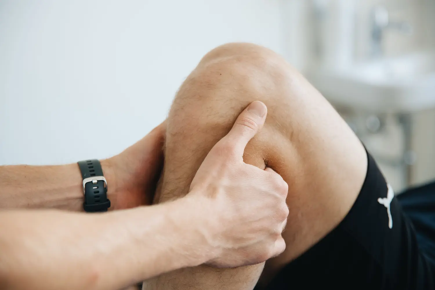
Close-up clinical examination of a flexed knee
“The question is not simply whether a tear exists—it is whether the tear explains the knee in front of you.”

CHAPTER 03 · IMAGING

MRI can show a meniscal tear.It cannot choose the treatment by itself.

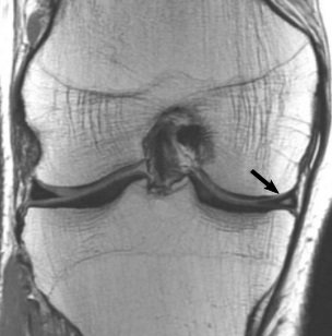
MRI is the preferred imaging test when an acute meniscal tear needs confirmation or when the result is likely to change management. In more gradual or degenerative presentations, imaging is interpreted alongside symptoms, examination and any arthritic change rather than treated as a diagnosis in isolation.

Proton-density coronal MRI of a knee showing a grade 2 medial meniscal tear, with an arrow identifying the abnormal meniscal signal
MRI: Lefevre et al.CC BY 4.0

SELECTED OBSERVATION 01 / 03

The MRI describes the tear pattern

Radiologists assess abnormal signal and morphology within the meniscus across multiple sequences and planes. The scan is read together with the history and examination rather than used as a stand-alone reason for surgery.

CHAPTER 04 · OPTIONS

Treatment follows the tear pattern.
Preserve useful meniscus when possible.

The route depends on whether the tear is acute or degenerative, whether the knee locks, the tear location and tissue quality, associated injuries, symptoms, function and the person’s goals.

ROUTE 01

SETTLE THE KNEE

Reduce irritation while protecting useful movement.

What it may include

Temporary activity modification, symptom control and a plan to maintain safe motion while swelling and pain settle.

What it cannot decide

Early symptom improvement does not reveal whether a tear is repairable or whether another structure is involved. Reassessment matters when symptoms persist.

See how the final decision is made

Not every tear follows the same route. Some settle with rehabilitation, some need further imaging, and selected symptomatic tears may be considered for surgery when the complete clinical picture supports it.

CHAPTER 05 · DECISION

When does meniscus surgery enter the conversation?

Not because an MRI reports a tear. Surgery becomes relevant when symptoms, examination, function and tear characteristics support a mechanical problem that is likely to benefit from an operative approach, or when an acute repairable tear is clinically suitable for preservation.

01

The tear fits the symptoms

The clinical pattern and examination support the meniscus as an important pain or mechanical source.

02

Function is genuinely limited

Locking, recurrent catching, pain with load or inability to return to required activity has meaningful impact.

03

The tear is assessed for preservation

Location, pattern, tissue quality, chronicity and blood supply influence whether repair is feasible.

04

The whole knee is considered

Ligament injury, cartilage damage and osteoarthritis may change the expected benefit and the procedure chosen.

Active adult rebuilding single-leg knee strength during rehabilitation

THE FUNCTION THAT MATTERS

The target is a knee you can trust under load.

Success is not simply the disappearance of a scan finding. It may mean walking, squatting, working, changing direction in sport or returning to training without recurrent swelling, locking or loss of confidence.

  • Walking & stairs
  • Squatting & kneeling
  • Work & daily loading
  • Sport & direction change

RELATED CLINICAL ROUTES

A meniscus tear may not be the only finding.

Knee symptoms overlap. These routes help compare the meniscus with ligament, cartilage and arthritic patterns without assuming the scan explains everything.

MENISCUS INJURIES FAQ

Questions worth answering before treatment is chosen.

General information only. A diagnosis and personal recommendation require an individual clinical assessment.

Request an appointment
01What does the meniscus do?

The knee has medial and lateral menisci. These fibrocartilage structures help distribute load, contribute to stability and protect the articular surfaces during movement.

02Does every meniscus tear need surgery?

No. Treatment depends on the tear pattern, symptoms, timing, associated knee problems, function and whether the tissue is likely to be repairable. Many tears can be managed without surgery.

03Do I always need an MRI?

No. History and examination come first. MRI is the preferred imaging test when an acute meniscal tear needs imaging confirmation or when the result is likely to change management. Degenerative tears often do not require immediate MRI, particularly when there are no persistent mechanical symptoms.

04Can a meniscus tear heal without surgery?

Some tears—particularly selected peripheral tears with a better blood supply—may heal or become manageable without surgery. Healing potential depends on location, pattern, stability, tissue quality and the rest of the knee.

05When is meniscus repair considered?

When surgery is indicated, repair may be considered when the tear pattern, location and tissue quality provide a reasonable healing opportunity. Preserving functional meniscal tissue is preferred when feasible.

06Repair or partial meniscectomy—what is the difference?

Repair aims to preserve and heal the torn meniscus. Partial meniscectomy removes only unstable or irreparable torn tissue when surgery is otherwise appropriate. Rehabilitation and recovery differ between the procedures.

07When does a locked knee need prompt assessment?

A knee that becomes acutely locked and cannot fully straighten after injury needs prompt clinical assessment because displaced meniscal tissue or another mechanical problem may be blocking motion.


YOUR CLINICAL STORY

The scan matters. The mechanism and function matter too.

Bring the injury timeline, what movements trigger pain or catching, whether the knee swells or locks, previous imaging and the activities you want to return to. The consultation connects those pieces before the treatment conversation begins.

Prof. Dr. Sammy Hanna, consultant orthopaedic hip and knee surgeon

YOUR SPECIALIST

Prof. Dr. Sammy Hanna

Prof. Hanna is a UK board-certified consultant orthopaedic hip and knee surgeon at Healthpoint Hospital, Abu Dhabi. His clinical interests include arthroscopic surgery for sports and soft-tissue knee injuries alongside primary, complex and revision joint replacement, partial knee replacement, minimally invasive techniques and robotic-assisted hip and knee replacement.

Chat with us